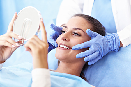
At Ramsi Dental, we take a thoughtful, preventive approach to oral health. Regular oral exams give our team the information we need to spot early signs of trouble, tailor care to each patient, and preserve long-term dental wellness. During routine visits we combine careful clinical assessment with patient education — from professional cleanings to screenings for oral cancer and periodontal concerns — so you leave with a clearer plan and greater confidence in your smile.
Your first comprehensive exam establishes a baseline: we review your medical history, discuss any symptoms or concerns, and listen to your oral health goals. That initial conversation helps us prioritize what to examine and which diagnostic tools to employ. We’ll ask about medications, past dental work, and habits such as grinding that can affect your teeth and jaw.
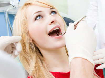
The clinical portion of the exam includes a systematic evaluation of teeth, gums, tongue, and the soft tissues of the mouth, as well as inspection of the head and neck for any unusual findings. We assess bite alignment and the function of the temporomandibular joints (TMJ), and we check for signs of wear, decay, infection, or inflammation. When appropriate, we recommend diagnostic imaging to see beyond what is visible to the naked eye.
Once the exam and any radiographs are complete, we’ll review findings with you in plain language and outline practical options for care. Whether the next step is a preventive cleaning, monitoring, or a treatment plan, our focus is on clear communication and shared decision-making so you understand your choices and feel comfortable moving forward.

Oral health is connected to overall health in ways many people don’t expect. Problems that start in the mouth — such as chronic gum inflammation or persistent infections — can influence other systems in the body. During an exam we look beyond teeth to identify signs that may warrant attention from your medical provider or further dental investigation.
Research increasingly shows links between oral conditions and systemic issues like cardiovascular disease, diabetes management, and respiratory health. While the relationships are complex, identifying and addressing oral disease early can play a role in reducing broader health risks. That’s why a thorough oral exam includes screening for tissue changes, persistent lesions, and other indicators that could signal a deeper concern.
We also track how medications, nutritional status, and lifestyle habits affect the mouth. Symptoms such as dry mouth, unusual bleeding, or persistent sores can result from non-dental causes; recognizing these signs allows us to coordinate care and provide appropriate referrals or recommendations to support your overall well-being.
Maintaining a healthy smile is a partnership between the care you give at home and the preventive services we provide in the office. Professional cleanings remove build-up in areas that are hard to reach with routine brushing and flossing, reducing the bacteria that cause cavities and gum disease. We tailor hygiene recommendations to each patient’s needs, offering practical tips to improve technique and motivation.
Preventive care also includes fluoride treatments, sealants when appropriate, and personalized guidance on diet and habits that protect enamel and gum tissue. For children, early preventive visits build good habits and let us monitor growth and jaw development. For adults, regular maintenance helps avoid complex problems later and preserves the natural dentition for as long as possible.
Our team recommends a schedule based on individual risk factors. Many patients benefit from twice-yearly checkups, while others with higher risk may need more frequent monitoring. The goal is to catch changes early, simplify treatment, and keep care as conservative and effective as possible.
Visual inspection is essential, but radiographs and other imaging tools reveal issues that may not be apparent on the surface. Dental x-rays help us detect decay between teeth, assess root and bone health, and identify developmental or structural concerns. When used judiciously, imaging is a safe and informative adjunct to the clinical exam.
Modern digital imaging reduces radiation exposure and provides images immediately, which improves the speed and clarity of diagnosis. These images are stored in the patient’s electronic record so we can compare them over time, track changes, and plan treatments with greater precision. We’ll explain why any image is recommended and how it informs your care.
For specific concerns — such as evaluating the position of impacted teeth, planning implant placement, or assessing complex anatomy — three-dimensional imaging like cone-beam CT may be recommended. This advanced view supports accurate treatment planning while helping us communicate options and anticipated outcomes clearly.
Diagnostic imaging comes in several forms, each suited to a particular clinical question. Small intraoral films, such as periapical and bitewing images, capture detailed views of individual teeth or the spaces between teeth and are commonly used for routine assessments. A full-mouth series gives a comprehensive view when a complete evaluation is needed.
Periapical x-ray - Shows an entire tooth from crown to root and the adjacent bone, useful for detecting root problems and infections.
Bitewing x-ray - Focuses on the crowns of back teeth and is especially helpful for locating decay between teeth.
Panoramic film - Provides a broad, two-dimensional overview of both jaws, useful for evaluating growth, impacted teeth, and general jaw health.
Cephalometric film - Offers a profile view used in orthodontic assessment and planning for skeletal relationships.
When three-dimensional detail is required, cone-beam computed tomography (CBCT) creates accurate 3D reconstructions that enhance diagnosis and surgical planning. While not needed for every patient, CBCT is a valuable tool in complex cases such as implant placement, evaluation of traumatic injuries, or detailed assessment of anatomical structures.
Routine oral exams are the foundation of lasting dental health: they identify issues early, guide preventive measures, and shape treatment plans that respect your goals and lifestyle. At Ramsi Dental, our team aims to make each exam informative, comfortable, and focused on long-term outcomes. If you have questions about what an exam involves or whether you should schedule an appointment, please contact us for more information.

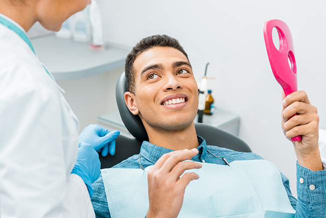
An oral exam is a comprehensive, preventive evaluation of the mouth and surrounding structures that helps identify early signs of disease and functional concerns. At Ramsi Dental, we combine a clinical inspection with a review of medical history and risk factors to create a clear picture of a patient’s oral health. The goal of an exam is to detect changes early, guide preventive care, and set priorities for any necessary treatment.
Exams typically include screening for cavities, gum disease, tissue changes and problems with bite or jaw function, and may involve targeted diagnostic tests when indicated. Findings are explained in plain language and used to develop a personalized plan that aligns with a patient’s goals and overall health. By emphasizing prevention and education, exams help patients maintain long-term oral wellness.
Your first comprehensive exam begins with a review of your medical and dental history, current medications and any symptoms or concerns you want to address. We will ask about habits such as grinding or tobacco use that affect oral health and listen to your goals so we can prioritize the exam. This conversation guides which diagnostic tools and tests are most appropriate for your care.
The clinical portion includes a systematic assessment of teeth, gums, tongue and other soft tissues, plus an inspection of the head and neck and an evaluation of bite and TMJ function. If indicated, we will take radiographs or other images to reveal issues below the surface. After the exam we review findings, discuss options and agree on a next step—whether that is preventive care, monitoring or a treatment plan.
Oral health and overall health are closely connected; conditions that begin in the mouth can affect other systems and, conversely, systemic diseases can show signs in oral tissues. Chronic gum inflammation and persistent infections have been associated with impacts on cardiovascular health, diabetes management and respiratory conditions, so identifying oral disease early can support broader health goals. During exams we look for tissue changes or persistent problems that may warrant medical follow-up.
We also consider how medications, nutrition and lifestyle influence oral symptoms such as dry mouth, unusual bleeding or delayed healing. Recognizing these signs allows us to coordinate care with a patient’s medical providers and provide practical recommendations that support both oral and systemic health. Clear documentation and timely communication help patients and their healthcare teams take appropriate next steps when needed.
Frequency of oral exams is individualized based on a patient’s risk factors, current oral health and medical history. Many patients benefit from twice-yearly checkups and cleanings, while people with higher risk for decay or gum disease—such as those with dry mouth, diabetes or a history of periodontal disease—may need more frequent visits. Your clinician will recommend an interval that balances prevention with efficient monitoring.
Regular exams focus on catching small changes before they become complex problems and on reinforcing home care habits that protect teeth and gums. A risk-based schedule helps keep treatments conservative and predictable while preserving natural dentition and oral function for as long as possible. We review the rationale for any recommended interval so patients understand the benefits of ongoing care.
Dental x-rays are a common and useful adjunct to the visual exam because they reveal decay between teeth, root and bone health, and other issues not visible on the surface. We use imaging judiciously, ordering only the views necessary to answer specific clinical questions and guide safe, effective care. Modern digital radiography reduces radiation exposure and produces images quickly for immediate review.
Images become part of the patient record so clinicians can compare them over time and track changes, which improves diagnostic accuracy and treatment planning. When three-dimensional detail is required for complex cases—such as implant planning or assessment of impacted teeth—cone-beam CT may be recommended. We always explain why any image is suggested and how it will influence the recommended care.
Oral cancer screening is an integral part of a thorough exam and includes careful visual and tactile inspection of the lips, tongue, oral mucosa, floor of the mouth and surrounding tissues. Clinicians look for persistent sores, red or white patches, lumps, unexplained bleeding or areas that do not heal, and they assess nearby lymph nodes for enlargement. Risk factors such as tobacco use, alcohol consumption and certain viral exposures are reviewed to inform the level of concern.
If a suspicious area is identified, we document its characteristics, monitor changes closely and, when appropriate, arrange for further evaluation or referral for biopsy. Early detection greatly improves outcomes, so timely follow-up and clear communication about next steps are emphasized. Patients are encouraged to report new or persistent symptoms between visits so lesions can be assessed promptly.
Before your appointment, assemble a list of current medications, recent medical history and any symptoms or questions you want to discuss. If you have recent dental records or x-rays from another provider, bringing them can reduce duplicate imaging and provide valuable historical context. It is also helpful to note habits such as clenching or tobacco use so the clinician can address potential risk factors.
On the day of the exam, continue your normal oral hygiene routine and arrive with any removable appliances or orthodontic devices you use. Be prepared to share changes in overall health, allergies or new medications since your last dental visit. Clear communication helps clinicians tailor the exam and make efficient, evidence-based recommendations.
Oral exams for children focus on growth, eruption patterns, preventive care and the development of healthy habits. Clinicians monitor tooth emergence and jaw development, assess for early signs of decay and discuss fluoride, sealants and nutrition to protect enamel. Behavior management and age-appropriate education are part of the visit so young patients become comfortable and engaged in their own care.
For adolescents, exams also evaluate changes in bite, wisdom tooth development and habits such as sports-related risks or tobacco use. Preventive strategies are adjusted as growth progresses and treatment needs evolve, and clinicians provide guidance to parents and caregivers on promoting long-term oral health. Regular pediatric-focused exams help avoid complex problems later and support healthy dental development.
Several common findings can alter a recommended plan, including new or progressing cavities, signs of periodontal disease such as pocketing or bleeding, significant occlusal wear or changes in jaw function. Soft-tissue abnormalities, persistent lesions or unexplained pain may prompt additional testing or referral to a specialist. Each finding is weighed against the patient’s goals and health status to determine the most appropriate path forward.
Options after an exam often range from enhanced preventive measures and closer monitoring to restorative or periodontal treatment when needed. The emphasis is on choosing conservative, evidence-based care that preserves oral structures and function whenever possible. We discuss alternatives, expected outcomes and follow-up so patients can make informed decisions about their care.
Diagnostic imaging provides detailed information about tooth roots, bone levels, anatomical relationships and structures that are not visible during a clinical exam alone. Two-dimensional images like bitewings and panoramic films are useful for routine diagnostics, while three-dimensional imaging delivers precise spatial detail for complex procedures such as implant placement or assessment of traumatic injuries. Having clear images helps clinicians identify the full scope of a problem and choose the most appropriate, targeted treatment.
Stored images also allow comparison over time, which aids in monitoring disease progression and evaluating treatment outcomes. Imaging enhances communication with patients by making findings more understandable and by supporting shared decision-making. When an image is recommended, we explain how it will inform care and how it contributes to a predictable, safe treatment approach.

We’d love to hear from you! Whether you have questions about our services, want to schedule an appointment, need guidance on your dental care, or simply want to learn more about how we can help you achieve a healthy, confident smile, our friendly and knowledgeable team is here to assist you.
Visit us at Ramsi Dental or reach out by phone or through our online form. We’re committed to making your experience easy, welcoming, and stress-free, and we’ll respond promptly to ensure you get the care you need.