Oral cancer can start quietly and progress without obvious symptoms, which is why routine screening during dental visits matters. Early detection dramatically improves the chances of successful treatment because abnormalities caught at an early stage are easier to evaluate and manage. Screening is a proactive step that complements good daily oral hygiene and regular care, serving as an important safety net for people of all ages.
A screening is not a diagnosis; it’s an examination that helps identify areas that may need further testing. Dentists look for visible changes in color, texture, and symmetry in the mouth and surrounding tissues, and when anything unusual appears, they recommend appropriate next steps. Because oral tissues heal and change over time, what looks normal one year can warrant attention the next — regular checks let clinicians track those changes.
Beyond clinical benefits, routine screening helps normalize conversations about symptoms and risk factors. Patients who know what to watch for and who receive clear information from their dental team are more likely to report concerns early. That collaborative approach between patient and provider supports better outcomes and gives people confidence that their oral health is being monitored comprehensively.
Certain factors increase the likelihood of developing oral cancer, but no single profile captures everyone at risk. Traditional contributors include tobacco use and frequent heavy alcohol consumption; together these behaviors have a stronger effect than either alone. Age and sex also influence risk patterns, with incidence rising in older adults and historically affecting men more often, though trends are changing over time.
Emerging risk factors have broadened the picture. Human papillomavirus (HPV), particularly high-risk strains, has been linked to cancers of the oropharynx and tonsils, shifting the demographic toward younger patients in some cases. Environmental exposures, past radiation therapy to the head and neck, and certain medical conditions that affect the immune system may also play a role. Eating a nutrient-poor diet and chronic irritation from ill-fitting dentures or rough teeth surfaces can contribute as well.
Risk assessment is an individualized process. A routine screening gives clinicians the chance to review a patient’s health history, lifestyle, and any new symptoms in context. That personalized perspective helps prioritize which findings warrant closer observation or referral to a specialist for diagnostic testing.
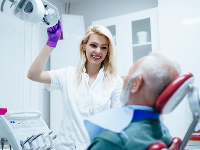
Oral cancer screening is quick, noninvasive, and typically performed as part of a comprehensive dental exam. The clinician begins with a conversation about your medical and dental history and asks about any new symptoms, such as persistent mouth sores, lumps or difficulty swallowing. This dialogue helps guide the visual and tactile inspection that follows.
During the exam, the dentist or hygienist will carefully examine the lips, tongue, cheeks, floor and roof of the mouth, gums, and the oropharynx. They use good lighting and may gently palpate the tissues and neck to feel for lumps or abnormalities beneath the surface. Any discoloration, ulcerations that do not heal, or unusual thicknesses are noted and measured against past records when available.
If a suspicious area is found, the clinician will explain what was observed and recommend the next steps. Those may include monitoring the site over time, documenting images, performing additional in-office tests, or referring to an oral surgeon or ENT specialist for biopsy and definitive diagnosis. The goal is to pursue clarity efficiently while avoiding unnecessary procedures.
In addition to a visual and tactile exam, dental practices may use adjunctive technologies designed to aid in early detection. Optical devices using specialized light or fluorescence can help highlight tissue changes that are not always evident under regular lighting. These tools are meant to support the clinician’s judgment, not replace a careful clinical exam.
Cytology brushes and oral swab tests provide another layer of evaluation by collecting cells from suspicious areas for laboratory analysis. When the clinical picture warrants it, a biopsy remains the gold standard for diagnosing oral cancer because it provides tissue for microscopic examination. Imaging studies, such as specialized scans, may be used by specialists to assess the extent of disease if a diagnosis is confirmed.
Good clinical practice combines technology with experience. Effective screening programs rely on trained clinicians who interpret findings in the context of each patient’s history and risk profile. This blended approach increases the likelihood of identifying clinically significant lesions while minimizing unnecessary alarm for benign conditions.
When a screening finds no concerns, patients benefit from reassurance and the knowledge that baseline observations are recorded for future comparison. Preventive care continues to be important: avoiding tobacco, moderating alcohol use, protecting lips from excessive UV exposure, eating a balanced diet, and maintaining regular dental visits all reduce risk and support overall oral health.
If follow-up is recommended, the dental team will outline a clear plan — whether that means a short-term recheck, further diagnostic testing, or referral to a specialist. Timely action and clear communication are essential components of effective care. Patients should feel empowered to ask questions about what was found, why a test is recommended, and what to expect next.
Raising awareness and fostering routine screening as part of dental care is a practical way to improve early detection rates. The team at Ramsi Dental emphasizes education and careful monitoring so patients understand both the process and the reasons behind recommended follow-up, ensuring appointments serve as meaningful checkpoints for oral health.
Oral cancer screening is a simple, routine procedure with the potential to make a profound difference in outcomes when abnormalities are found early. By combining careful clinical exams with appropriate use of adjunctive tools and individualized follow-up plans, dental teams can help detect problems at a stage when treatment is most effective.
If you have questions about what to expect during a screening, have noticed any persistent changes in your mouth, or would like to make oral cancer screening a regular part of your dental care, please contact us for more information. Our goal is to provide clear, compassionate guidance and to support you in maintaining long-term oral health.


An oral cancer screening is a focused clinical exam that evaluates the lips, tongue, cheeks, floor and roof of the mouth, gums and the oropharynx for unusual signs or symptoms. It is a noninvasive check performed during a routine dental visit to identify abnormalities early, when they are most treatable. The screening does not provide a diagnosis but helps determine whether further testing or specialist referral is needed.
The process typically combines visual inspection with gentle palpation of tissues and the neck to feel for lumps or thickened areas. Clinicians review medical and social history as part of the assessment to place findings in context. Results are documented so changes can be tracked at future visits.
Routine screening increases the chance of detecting precancerous or early-stage cancers before symptoms become severe, which can significantly improve treatment outcomes. Early detection often allows for less extensive treatment and better preservation of oral functions. Regular screenings also establish a record of what is normal for each patient, making it easier to spot subtle changes over time.
Screenings normalize conversations about symptoms and risk factors so patients know what to monitor between visits. They also enable clinicians to provide personalized prevention advice informed by a patient’s history and habits. Together, these measures support timely evaluation and coordinated care when needed.
Most adults benefit from an oral cancer screening at least once a year as part of their comprehensive dental exam, since risk increases with age and cumulative exposures. People with known risk factors—such as tobacco use, heavy alcohol consumption, a history of head and neck radiation, or immune suppression—may need more frequent checks. Emerging factors, including high-risk strains of HPV, have broadened the groups that should be monitored closely.
Screening frequency should be individualized based on medical history and clinical findings, so patients with suspicious lesions or changing symptoms will typically be seen more often. Your dental clinician will recommend an appropriate schedule and explain the rationale for closer follow-up when indicated. Proactive scheduling helps ensure meaningful continuity of care.
The clinician begins by asking about medical history and any new symptoms, such as persistent sores, lumps or difficulty swallowing, to guide the exam. The visual inspection uses good lighting to examine soft tissues, while gentle palpation of the mouth and neck checks for hidden masses. Any areas of discoloration, ulceration or unusual texture are measured and compared with prior records when available.
If something of concern is identified, the clinician will explain observations, document the findings with notes or images, and outline recommended next steps. Those steps can include short-term monitoring, adjunctive testing, or referral for biopsy to establish a definitive diagnosis. Clear communication ensures patients understand what was found and why further action may be necessary.
Patients should seek evaluation for persistent mouth sores, unexplained lumps or lumps that do not resolve, areas of white or red patching, numbness, or difficulty chewing and swallowing. Unexplained bleeding or loose teeth without an obvious cause, and changes in voice or a chronic sore throat, can also be warning signs. Any oral changes that last more than two weeks warrant prompt attention.
Early reporting of symptoms improves the chance of timely diagnosis and treatment, so patients are encouraged to contact their dental team promptly when they notice changes. The dental clinician will assess the symptom in context and determine whether immediate testing or a short-interval recheck is appropriate. Documenting symptoms and their progression helps guide clinical decisions.
Adjunctive technologies such as fluorescence or specialized light-based devices can help highlight abnormal tissue that may be less visible under standard lighting, acting as a supplement to the clinical exam. Cytology brushes and oral swab tests collect cells from suspicious sites for laboratory analysis that can aid in triage. While these tools add information, they do not replace the clinical judgment of a trained examiner.
A biopsy remains the definitive test when tissue diagnosis is required, providing material for microscopic examination and accurate staging when cancer is present. Imaging studies may be used by specialists to assess the extent of disease after diagnosis. Effective programs use technology selectively and interpret results in light of each patient’s history and exam findings.
If a suspicious lesion is detected, the clinician will explain the finding, answer questions, and recommend a clear follow-up plan tailored to the level of concern. Options commonly include short-term monitoring with documented images, adjunctive testing, or referral to an oral surgeon or ENT specialist for biopsy. The goal is to obtain diagnostic clarity while avoiding unnecessary invasive procedures when a lesion appears benign.
When a biopsy or specialist evaluation is advised, the dental team will coordinate referrals and share relevant records to support continuity of care. Timely communication and documentation help streamline the diagnostic process and reduce delays. Patients should feel empowered to ask about the reasons for each recommended step and what to expect from the next appointment.
Reducing risk involves lifestyle choices and preventive care, including avoiding tobacco in all forms, limiting alcohol consumption, maintaining a balanced diet rich in fruits and vegetables, and protecting lips from excessive sun exposure. Addressing chronic irritation from ill-fitting dentures or sharp tooth edges and managing conditions that affect immune function also lowers risk. Regular dental visits for screening and early intervention are a key component of risk reduction.
Vaccination against HPV can reduce the likelihood of cancers linked to high-risk HPV strains and is an important public health measure for eligible individuals. Clinicians can help patients understand risk factors and create realistic, individualized strategies to minimize exposure. Education and routine monitoring together support long-term oral health.
High-risk strains of human papillomavirus (HPV) have been linked to cancers of the oropharynx and tonsils, and their association has changed the epidemiology of head and neck cancers in recent years. HPV-related cancers often present differently and may occur in younger patients who lack traditional tobacco- or alcohol-related risk factors. Assessment includes a thorough history, targeted clinical exam, and, when appropriate, referral for further testing or specialist evaluation.
Vaccination and education are central to prevention efforts for HPV-associated disease, and clinicians discuss these options with patients as part of comprehensive care. When a lesion is suspicious in an area commonly affected by HPV-related cancers, the dental team may coordinate testing and specialist referral to establish the underlying cause. Early detection remains critical regardless of the etiologic factor.
Ramsi Dental emphasizes routine, evidence-based screening as part of comprehensive dental care, combining careful visual and tactile exams with selective use of adjunctive technologies when clinically indicated. The practice records baseline observations and communicates findings clearly so patients understand what was noted and why a particular follow-up plan is recommended. Clinicians tailor monitoring intervals and referrals to each patient’s risk profile and clinical picture.
When further evaluation is necessary, Ramsi Dental coordinates referrals and provides documentation to specialists to support timely diagnosis and treatment planning. The team prioritizes patient education and clear communication so individuals know what to watch for between visits and when to seek care. This structured approach helps ensure that screening appointments serve as reliable checkpoints for long-term oral health.

We’d love to hear from you! Whether you have questions about our services, want to schedule an appointment, need guidance on your dental care, or simply want to learn more about how we can help you achieve a healthy, confident smile, our friendly and knowledgeable team is here to assist you.
Visit us at Ramsi Dental or reach out by phone or through our online form. We’re committed to making your experience easy, welcoming, and stress-free, and we’ll respond promptly to ensure you get the care you need.