An intraoral camera is a compact, pen-sized imaging device designed to capture detailed, full-color views of the teeth and surrounding soft tissues. Unlike an external camera, it is purpose-built for use inside the mouth, delivering high-resolution photos and live video that are immediately viewable on a monitor. The result is a clear, magnified perspective that helps both clinicians and patients see areas that are otherwise difficult to visualize.
These cameras use bright LED lighting and close-focus optics to reveal surface texture, cracks, restorations, and areas of inflammation with clarity. The images are typically displayed in real time so the dental team can review them with the patient, highlight points of concern, and explain recommended options. Because the device sits just millimeters from the tooth surface, it can capture angles and details that traditional mirrors or written descriptions cannot.
Image capture is quick and noninvasive; most examinations take only a few seconds per view. The technology complements traditional exam techniques rather than replacing them, providing an additional diagnostic perspective that supports better-informed care decisions. For patients, seeing a crisp image often makes oral health issues easier to understand and remember.
One of the primary advantages of intraoral cameras is their ability to turn abstract dental concepts into concrete visuals. Rather than relying solely on verbal descriptions, clinicians can show patients exactly what they see: a worn filling, early tooth decay, a hairline crack, or an area of gum irritation. This visual evidence fosters clearer conversations and helps patients grasp the condition of their mouth at a glance.
Seeing an image on screen allows patients to participate in treatment planning more actively. When problems are visible, questions become more focused and productive, and patients are better able to weigh the pros and cons of different approaches. This transparency also supports informed consent because patients can review the same images the dental team uses to form recommendations.
In everyday practice, intraoral photos are an excellent educational tool for both routine check-ups and more complex discussions. They can be used to demonstrate proper cleaning techniques, show changes over time, and set realistic expectations for restorative or cosmetic procedures. For many patients, that combination of visual learning and professional explanation reduces anxiety and increases confidence in their care plan.
Clinically, intraoral cameras are versatile instruments that support a range of diagnostic tasks. They help detect early decay that might not be obvious on X-rays, evaluate the margins of existing restorations, and reveal soft-tissue conditions such as ulcers or lesions. While not a substitute for radiographs or clinical probing, intraoral imaging provides complementary information that often changes or clarifies clinical decisions.
For treatment planning, captured images become part of the patient record and can be referenced when discussing restorative, periodontal, or cosmetic options. Clear photographs help clinicians plan procedures with greater precision and track progress after treatment. Because images are dated and stored, they serve as a reliable baseline for future comparisons and can reveal subtle changes that might otherwise go unnoticed.
Accurate documentation is also important for collaboration with specialists, laboratories, and other members of the dental team. High-quality images can be shared with a prosthodontist, endodontist, or oral surgeon to provide additional context before a referral. They are likewise useful when communicating with dental laboratories to ensure shade, contour, and margin expectations are understood.
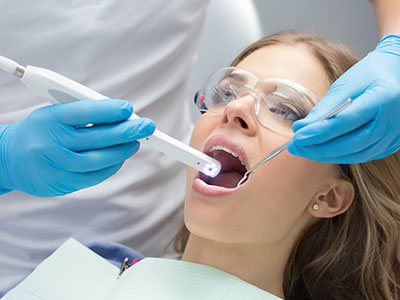
An intraoral imaging session is typically comfortable and quick. The clinician or hygienist will guide the camera into the mouth, capturing the views needed to diagnose or document a condition. Because the device is small and the lighting is integrated, the procedure is less intrusive than some older imaging techniques and is generally well tolerated by patients of all ages.
During the process, images appear on a monitor in real time so the clinician can point out findings and explain their significance. Patients are encouraged to ask questions as they view the images; this shared review often makes treatment goals and timelines clearer. If additional images are needed later, the clinician can take them during routine visits to monitor changes over time.
Because digital files are created instantly, there is no delay in adding photos to the patient chart. The speed of capture minimizes chair time while maximizing the detail available for diagnosis. For patients, this means a more informative visit without a significant increase in appointment length.
Modern intraoral cameras connect seamlessly with dental practice management and imaging software, allowing photos to be labeled, archived, and retrieved quickly. Digital storage safeguards patient images as part of the permanent record and enables clinicians to compare current photos with past visits to detect progression or improvement. Many practices use secure, HIPAA-compliant systems to maintain privacy and accessibility.
These images are also valuable when coordinating care with outside providers. A well-documented photo package sent to a specialist or dental laboratory helps ensure everyone involved has the same visual reference. This reduces miscommunication and improves the precision of referrals, lab prescriptions, and interdisciplinary treatment plans.
As imaging technology continues to advance, intraoral cameras are becoming more integrated with other diagnostic tools—such as digital impressions and cone-beam imaging—so clinicians can assemble a comprehensive picture of a patient’s oral health. The practice’s adoption of these tools demonstrates a commitment to modern, patient-centered care and a focus on accuracy and transparency.
In summary, intraoral cameras bring clarity, efficiency, and collaboration to routine dental care. They enhance communication between clinician and patient, support informed treatment decisions, and provide durable documentation for ongoing care. If you’d like to learn more about how intraoral imaging is used in our office or how it might benefit your next visit, please contact us for more information.


An intraoral camera is a small, pen-sized device that captures high-resolution, full-color images and live video of the teeth and surrounding soft tissues. It uses close-focus optics and bright LED illumination to reveal surface texture, cracks, restorations, and areas of inflammation that are difficult to see with the naked eye. Images appear on a monitor in real time so clinicians and patients can review findings together during the appointment.
The camera sits just millimeters from the tooth surface, allowing it to record angles and details that traditional mirrors and verbal descriptions cannot convey. Image capture is quick and noninvasive, typically taking only a few seconds per view. Because files are digital, photos can be archived immediately in the patient record for future comparison and documentation.
Intraoral images turn abstract descriptions into concrete visuals, helping patients understand exactly what the clinician sees. Showing a worn filling, early decay, or gum irritation on a screen focuses the conversation and makes treatment options clearer. This shared visual reference encourages more specific questions and supports informed decision making.
Viewing images together also helps set realistic expectations for restorative and cosmetic procedures by demonstrating existing conditions and likely outcomes. For many patients, seeing their own oral condition reduces uncertainty and strengthens trust in the diagnostic process. Clinicians can use images as teaching tools to show proper home care techniques and changes over time.
Clinically, intraoral cameras provide complementary information to radiographs and clinical probing by exposing surface details that X-rays may miss. They help detect early enamel breakdown, evaluate the margins of existing restorations, and document soft-tissue abnormalities such as ulcers or lesions. These visual details can influence diagnosis and guide choices about preventive, restorative, or periodontal care.
Captured images become part of the permanent record and support precise treatment planning by allowing clinicians to reference dated photographs when preparing procedures. High-quality images are useful for collaboration with specialists and dental laboratories, providing a consistent visual reference for referrals and lab prescriptions. Regular photographic documentation also helps clinicians monitor healing and long-term outcomes.
An intraoral imaging session is generally quick, comfortable, and minimally intrusive. The clinician or hygienist will guide the camera into the mouth and capture the necessary views while the patient remains seated; integrated lighting and small optics make the device easy to tolerate. Images are displayed on a monitor immediately so the clinician can explain findings as they appear.
The realtime review encourages questions and clarifies treatment goals without significantly extending appointment length. If follow-up photos are needed, they can be taken during routine visits to track changes. Digital capture also reduces chair time associated with developing or processing images.
Yes, intraoral cameras are designed to be safe and comfortable for patients of most ages. The devices are small and lightweight, with rounded tips to minimize gagging or irritation, and they require no radiation or invasive procedures to obtain images. Clinicians adjust the approach to accommodate children, anxious patients, or those with a strong gag reflex.
Because imaging is noninvasive and quick, it is usually well tolerated and can be introduced gradually to build patient comfort. Infection control protocols are followed closely, including barrier sheaths and proper sanitization between uses, to maintain a safe environment. If a patient has concerns, the dental team can explain the process and proceed at a comfortable pace.
Modern intraoral images are captured as digital files and integrated with practice management and imaging software for convenient archiving. These systems label and store images with visit dates and patient identifiers so clinicians can easily retrieve past photos for comparison. Many practices use HIPAA-compliant platforms to ensure that images remain private and accessible only to authorized team members.
Stored images serve as reliable baseline documentation and facilitate continuity of care across providers. When images are shared with specialists or laboratories, they are transmitted securely to preserve patient confidentiality. Proper digital management also simplifies tracking of treatment progress and long-term monitoring.
Intraoral cameras increasingly integrate with a range of diagnostic and restorative technologies to create a more complete clinical picture. Images can be combined with digital impressions, radiographs, and cone-beam scans to inform comprehensive treatment planning. This interoperability helps clinicians correlate surface detail with subsurface anatomy and plan procedures with greater precision.
Integration also streamlines communication with laboratories and specialists by supplying consistent visual references alongside other diagnostic data. As imaging technology evolves, integration enhances workflow efficiency and supports a patient-centered approach to care. Practices that adopt interconnected systems can offer more coordinated and predictable outcomes.
While intraoral cameras excel at capturing surface detail and enhancing patient communication, they do not replace radiographs or clinical probing for many diagnostic purposes. Internal structures, bone levels, and interproximal decay are often better evaluated with X-rays or three-dimensional imaging. Intraoral photos should be viewed as a complementary tool that augments, rather than substitutes for, other diagnostic methods.
Some limitations include restricted depth perception and difficulty imaging posterior surfaces in patients with a limited mouth opening. Image quality can also be affected by moisture, reflections, or patient movement. Clinicians combine photographic data with clinical examination and imaging studies to form a complete and accurate diagnosis.
At Ramsi Dental, intraoral images are used to document current conditions, explain findings to patients, and support treatment planning discussions. Clinicians review images with patients during the visit to highlight areas of concern and outline possible approaches to care. Photographs are archived in the patient record to track changes and evaluate treatment results over time.
Images are also shared securely with specialists or dental laboratories when collaborative care is needed, ensuring everyone involved has the same visual reference. This coordinated approach improves clarity in referrals and helps the team deliver more precise restorative and cosmetic outcomes. Patients are encouraged to ask questions as images are reviewed to better understand their oral health and care options.
In most cases, no special preparation is required for intraoral imaging beyond routine oral hygiene. Patients should simply arrive with their mouth clean; clinicians may remove excess saliva or debris during the session to capture clear views. If a particular area needs to be examined in detail, the clinician will give brief, specific instructions during the appointment.
If imaging is part of a more extensive diagnostic visit, the dental team will explain any additional steps needed in advance. Otherwise, the quick and noninvasive nature of intraoral photography makes it easy to include as part of routine checkups and treatment planning visits. Patients with specific concerns can mention them at check-in so the clinician can allocate appropriate time for imaging.

We’d love to hear from you! Whether you have questions about our services, want to schedule an appointment, need guidance on your dental care, or simply want to learn more about how we can help you achieve a healthy, confident smile, our friendly and knowledgeable team is here to assist you.
Visit us at Ramsi Dental or reach out by phone or through our online form. We’re committed to making your experience easy, welcoming, and stress-free, and we’ll respond promptly to ensure you get the care you need.