Digital radiography uses electronic sensors and computer systems to capture dental X-ray images, replacing the older chemical film process. The image is recorded on a digital sensor and transmitted instantly to a computer, where it can be reviewed, enhanced, and stored with the patient’s records. This shift from film to digital changes how clinicians diagnose, plan, and communicate about dental care.
For patients, the most noticeable changes are speed and convenience: images appear immediately, removing the wait time associated with film development. Clinicians can adjust brightness, contrast, and magnification in real time, which helps reveal subtle details without taking additional exposures. The immediate feedback also supports more efficient appointments and clearer conversations about treatment options.
Beyond convenience, digital radiography supports a more coordinated workflow. Files are stored electronically and can be shared securely between providers when additional opinions or referrals are needed. That interoperability helps maintain continuity of care across specialists, laboratories, and other practices.
Ramsi Dental uses digital radiography to streamline diagnostics while keeping patient comfort and safety at the forefront. The technology allows our team to work more efficiently and spend more time discussing findings rather than waiting for images to develop.
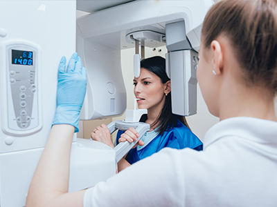
The procedure for a digital dental X-ray is similar to traditional X-rays but with a few modern differences. A small sensor is placed inside or near the mouth in the area being examined; the patient is positioned and asked to remain still while the image is captured. The exposure itself is very quick—often just a fraction of a second—and the result is sent instantly to a computer monitor.
Because images are immediate, the clinician can confirm they have captured the required view without delay, reducing the need for repeat exposures. For many patients this translates into shorter appointment times and less time spent repositioning or re-taking images. The sensor is designed to be thin and as comfortable as possible, and staff are trained to minimize discomfort during placement.
Digital systems also allow for a variety of image types—bitewing, periapical, and panoramic—depending on clinical needs. The choice of sensor and technique is determined by the diagnostic question and the patient’s comfort, and the team will explain what type of image is needed before proceeding.
Throughout the process, infection-control protocols and equipment maintenance are followed to ensure safety and hygiene. If you have any concerns about positioning or comfort, the clinical team will address them immediately to make the experience as smooth as possible.
One of the primary benefits of digital radiography is improved image quality and the ability to manipulate images after capture. Clinicians can magnify areas of interest, adjust contrast, and apply filters that make small changes in tooth structure or bone more visible. These capabilities support earlier detection of cavities, fractures, and bone changes that might otherwise be harder to spot on conventional film.
Digital images also facilitate comparative review. Because images are stored with timestamps in electronic records, clinicians can quickly compare current images to earlier ones to monitor progression or healing over time. This longitudinal view is especially useful when tracking treatment outcomes or monitoring chronic conditions.
In addition to diagnostic clarity, digital radiography helps with treatment planning. Enhanced images improve the precision of measurements used for restorations, root canal work, and implant planning. When coordinated with other digital tools in the dental office, radiographs become part of an integrated clinical picture that supports predictable, evidence-based decisions.
Because fewer retakes are typically needed and image adjustments can reduce interpretation errors, digital radiography contributes to more consistent and reliable clinical documentation—benefits that matter both to clinicians and to patients who want informed recommendations.
Digital radiography generally requires less radiation than older film-based systems, thanks to more sensitive sensors and optimized exposure settings. Clinicians follow established safety practices—such as using appropriate shielding and exposure protocols—to minimize dose while obtaining diagnostic-quality images. Those practices are part of a standard approach to keep radiation exposure as low as reasonably achievable.
From a patient comfort perspective, the thin digital sensors used today are designed to be as unobtrusive as possible. The reduced need for repeated images also minimizes the time a sensor must remain in the mouth. Many patients find the overall experience quicker and less invasive than the film process used in the past.
There are environmental benefits as well: digital radiography eliminates the chemical processing required for film development and reduces paper and packaging waste. The electronic storage of images reduces the need for physical archives and supports a more sustainable clinical footprint.
Maintaining digital equipment and following best practices ensures these safety and environmental advantages are realized without sacrificing diagnostic effectiveness.
Digital radiographs are easily incorporated into electronic health records, which simplifies documentation and retrieval. Clinicians can tag and file images alongside notes, treatment plans, and other diagnostic data so that the entire clinical team has access to the same up-to-date information. This organized approach reduces administrative friction and supports coordinated care.
For patients, the ability to view their own images on screen during an appointment enhances understanding and engagement. Visual aids help clinicians explain diagnoses, illustrate the rationale for proposed treatments, and show progress over time. Seeing images side-by-side with annotated explanations makes clinical recommendations more transparent and easier to follow.
When collaboration with outside specialists or laboratories is needed, digital files can be shared securely and efficiently, speeding up referrals and second opinions. That accessibility promotes timely decision-making and helps ensure that all members of the care team have the information they need to act confidently.
Wrap-up: Digital radiography brings faster results, clearer imaging, and improved patient communication to modern dental care. If you’d like to learn more about how we use this technology in our practice, please contact us for more information. Ramsi Dental is happy to answer your questions and explain how digital imaging supports better diagnosis and treatment planning.


Digital radiography uses electronic sensors and computer systems to capture dental X-ray images instead of film. The sensor records an image that appears instantly on a computer where clinicians can view and enhance it for diagnosis. This technology speeds up appointments and allows clearer communication about findings.
At Ramsi Dental we use digital radiography to streamline diagnostics while prioritizing patient comfort and safety. Immediate image review reduces repeat exposures and gives our team more time to discuss treatment options with patients. Files are saved directly into the electronic record for long-term comparison and continuity of care.
Instead of chemical film, digital systems use flat-panel sensors or phosphor plates to capture X-rays, producing a digital file rather than a physical negative. Because images are delivered to a monitor instantly, clinicians can adjust brightness, contrast and magnification without re-taking films. The absence of darkroom chemicals and physical processing also reduces environmental impact.
Traditional film required longer processing and manual storage, while digital images integrate with electronic health records for easier access and comparison. Digital exposure settings and more sensitive sensors typically lower radiation dose compared with older film techniques. The result is a faster, more efficient workflow that supports detailed diagnosis.
Digital radiography generally uses lower radiation doses than conventional film X-rays because modern sensors are more sensitive and exposures are optimized. Dental teams follow the ALARA principle—keeping radiation as low as reasonably achievable—by selecting appropriate settings, using shielding and limiting images to those that are clinically necessary. Patients should always inform the clinical team if they are pregnant or believe they may be pregnant so staff can take additional precautions.
Protective measures commonly include lead aprons, thyroid collars when indicated and careful positioning to avoid repeat exposures. For children and vulnerable patients, clinicians tailor protocols and choose the least invasive imaging needed to answer the diagnostic question. These standard practices help ensure that diagnostic benefit outweighs any minimal risk.
Common intraoral digital images include bitewings, which assess decay between teeth, and periapical views that show the entire tooth and surrounding bone. Panoramic images capture the full arches and jaw relationships in a single image, useful for surgical planning and broad screening. Cone beam computed tomography (CBCT) provides three-dimensional data for complex implant planning, endodontic assessment or evaluation of anatomical structures.
The choice of image type depends on the clinical question, patient anatomy and the level of detail required for treatment. Clinicians will explain which view is needed and why, balancing diagnostic value with patient comfort. Modern digital suites allow these image types to be compared and integrated for comprehensive treatment planning.
In most cases no special preparation is required for digital dental X-rays, and patients can eat, take medications and arrive as usual for their appointment. Wearable metal such as necklaces or removable dental appliances may be asked to be taken out if they interfere with the image area. If you have a specific health condition or are pregnant, inform the staff ahead of time so the team can plan appropriately.
Children and nervous patients may benefit from a brief explanation of the steps so they know what to expect, and staff can provide extra reassurance during positioning. The clinician will position the sensor carefully and ask you to remain still for a moment while the image is acquired. Immediate review allows any needed adjustments to be made without multiple exposures.
Digital radiographs support diagnosis by allowing clinicians to magnify areas of interest, adjust contrast and apply filters that can reveal subtle decay, fractures or bone changes. These enhancements improve detection and help clinicians make timely, evidence-based recommendations. Timestamped images stored in the record enable direct comparison with prior studies to monitor progression or healing.
For treatment planning, precise measurements from digital images help with restorations, root canal work and implant placement by clarifying margins and anatomical landmarks. When combined with other digital tools, radiographs contribute to predictable surgical and restorative outcomes. Clear, annotated images also help clinicians explain diagnoses and options to patients, improving shared decision making.
Digital radiographs are stored electronically in secure practice management systems and integrated electronic health records, which simplifies retrieval and long-term archiving. Access controls, user authentication and routine backups are part of standard data management to protect patient information. When images are shared with outside specialists, secure methods such as encrypted file transfer or protected portals are used to maintain confidentiality.
Maintaining accurate metadata, including dates and source identifiers, ensures images remain linked to the correct patient record and supports clinical continuity. Regular software updates and adherence to privacy regulations are important to reduce security risks. Patients can request explanations about how their images are stored and with whom they may be shared as part of their care.
Because digital systems provide immediate feedback and higher initial image quality, the need for repeat X-rays is reduced compared with film, which could require reprocessing or retakes. Clinicians can confirm image completeness on the spot and make small adjustments electronically to enhance diagnostic detail. Fewer retakes mean shorter appointments and less cumulative radiation for patients.
Training and consistent positioning protocols for staff further decrease the likelihood of repeat exposures by ensuring reproducible, diagnostic views. When a repeat is unavoidable, technicians evaluate alternatives and use the lowest exposure necessary to obtain the needed information. This approach supports reliable documentation while minimizing unnecessary imaging.
Sensor design and proper positioning help maximize patient comfort during digital X-rays; most contemporary sensors are thin and contoured to reduce gagging or discomfort. Staff are trained to place sensors gently and to adjust techniques for patients with limited mouth opening or sensitive soft tissues. If a patient reports discomfort, the team will pause and modify the approach to improve comfort while maintaining image quality.
Infection control is maintained through barrier protection on intraoral sensors, strict instrument cleaning protocols and routine equipment maintenance. Disposable covers, surface disinfectants approved for dental equipment and staff adherence to standard precautions help keep imaging safe and hygienic. These measures are combined with routine quality checks to ensure images remain sharp and reliable.
Digital radiography streamlines coordination of care because high-quality images can be shared quickly with specialists, laboratories and referring providers, supporting timely consultations and treatment sequencing. Integrated digital files and accompanying notes help external clinicians understand the diagnostic context without waiting for physical films. This speed and clarity can improve the efficiency of multi-disciplinary care.
At Ramsi Dental we emphasize clear documentation and secure sharing practices so that referring clinicians receive the information they need to make informed recommendations. Digital tools also make it easier to include annotated images in referral packages, which reduces ambiguity and speeds decision making. The overall result is smoother collaboration and more cohesive patient care.

We’d love to hear from you! Whether you have questions about our services, want to schedule an appointment, need guidance on your dental care, or simply want to learn more about how we can help you achieve a healthy, confident smile, our friendly and knowledgeable team is here to assist you.
Visit us at Ramsi Dental or reach out by phone or through our online form. We’re committed to making your experience easy, welcoming, and stress-free, and we’ll respond promptly to ensure you get the care you need.