At the office of Ramsi Dental, we rely on advanced diagnostics to guide treatment decisions and deliver predictable results. Cone-beam computed tomography (CBCT) is one of the most valuable tools in our imaging suite: it captures high-resolution, three-dimensional views of teeth, jaws, and adjacent anatomy so clinicians can evaluate complex conditions with clarity and confidence.
Our practice uses a modern CBCT system that balances image quality with patient safety. The scans produce distortion-free, multi-planar views that reveal relationships and details conventional X-rays cannot, while allowing us to focus radiation only where it is clinically necessary.
Traditional dental X-rays provide important two-dimensional information, but they compress depth and can hide critical details. CBCT produces volumetric datasets that let clinicians look through slices of bone and tooth structure from any angle. This three-dimensional perspective exposes root configurations, cortical plates, sinus anatomy, and the position of nearby nerves with much greater clarity.
Because CBCT captures the spatial relationships between structures, it helps identify problems that often go undetected on flat films — for example, the course of a mandibular canal near a prospective implant site, a fractured root that is overlapped on a 2D image, or the full extent of a cyst or lesion. These insights lead to more informed decisions about diagnostics and treatment sequencing.
Importantly, CBCT is not a replacement for clinical examination or traditional films; it is a complementary modality. We use it selectively when three-dimensional detail will change diagnosis, risk assessment, or the proposed treatment plan.
CBCT equips clinicians with precise anatomical information that enhances diagnostic accuracy across multiple disciplines. Endodontists use it to locate additional canals or identify resorption; oral surgeons use it to evaluate impactions and pathologic conditions; orthodontists review airway and skeletal relationships to refine treatment strategy. In each case, the extra spatial detail minimizes surprises during treatment.
The volumetric data are also invaluable for planning. With CBCT, we can measure bone height and width, assess angulation, and determine the safest trajectory for surgical instruments or implants. These measurements are taken directly from the dataset rather than estimated from a flat image, reducing guesswork and improving reproducibility.
Beyond measurements, CBCT images are used to simulate procedures and to create guides or models when appropriate. That simulation capability supports collaborative case planning among specialists and helps set realistic expectations for outcomes.
For implant dentistry and oral surgery, CBCT has become a standard of care because it clarifies the anatomy that matters most for safety and success. The scans allow us to visualize bone density, detect undercuts, and evaluate proximity to vital structures such as the inferior alveolar nerve or the maxillary sinus—information that directly informs implant selection and placement strategy.
When a case warrants it, the CBCT dataset can be integrated with digital planning software to design surgical guides. These guides translate the virtual plan into a precise clinical execution, increasing accuracy and reducing chairside time. Even when guides are not used, preoperative CBCT review markedly improves intraoperative predictability.
Surgeons also rely on CBCT when assessing pathology or planning extractions of impacted teeth. The three-dimensional view helps anticipate complications, choose the most conservative access, and protect adjacent teeth and tissues during intervention.
Patient safety is a priority in every imaging decision. Modern dental CBCT units are engineered to limit exposure by using focused fields of view and optimized exposure settings tailored to the diagnostic task. When used appropriately, CBCT delivers a lower radiation burden than medical CT scans and concentrates imaging to the area of interest rather than exposing the whole head and neck.
Our team follows established radiation-safety principles, including the ALARA concept (as low as reasonably achievable), selecting the smallest field of view and lowest dose that will still yield diagnostic-quality images. We also screen every patient to confirm that a CBCT scan is truly indicated and that the expected benefit outweighs any exposure.
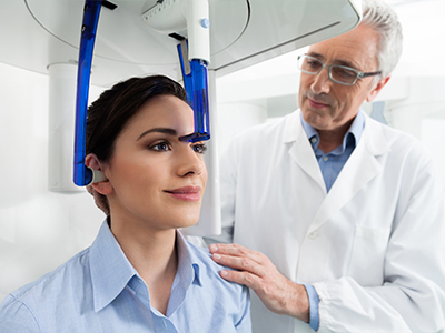
In terms of comfort, CBCT exams are noninvasive and typically completed within a minute or two. Patients remain seated or standing while the scanner rotates around the head, and there is no need for intravenous contrasts or injections. This quick, open-architecture design reduces anxiety and improves compliance, especially for those who find traditional medical imaging uncomfortable.
The value of CBCT lies in how the information is applied. At our practice, imaging findings are combined with clinical evaluation, patient history, and restorative goals to develop individualized treatment plans. That integrated approach ensures that CBCT contributes directly to decisions about timing, technique, and the level of intervention required.
We also use three-dimensional visualizations to help patients understand their situation. When appropriate, clinicians review axial, coronal, and sagittal views with patients so they can see the same information that guided the clinical recommendation. This transparency supports shared decision-making and helps patients feel more confident about next steps.
Finally, CBCT datasets facilitate collaboration with specialists when cases are complex. Digital files can be reviewed by multidisciplinary teams to coordinate care, reduce redundant imaging, and streamline treatment. This interconnected workflow improves efficiency and often leads to better outcomes.
In summary, cone-beam computed tomography is a powerful diagnostic tool that enhances visualization, supports precise planning, and contributes to safer, more predictable dental care. To learn more about how we use CBCT in diagnosis and treatment planning, or to discuss whether it is appropriate for your situation, please contact us for more information.


Cone-beam computed tomography (CBCT) is a three-dimensional imaging modality designed for dental and maxillofacial applications. It captures a volumetric dataset by rotating a cone-shaped X-ray beam around the patient, producing images in axial, coronal and sagittal planes. The result is a high-resolution view of teeth, bone and surrounding anatomy that supports detailed assessment.
CBCT differs from medical CT in geometry, dose management and dental-focused software tools, which make it practical for office-based imaging. The datasets can be reviewed as individual slices or rendered into reformatted views to visualize complex relationships. This capability makes CBCT a valuable adjunct to clinical examination when three-dimensional detail is needed.
Conventional dental X-rays produce two-dimensional projections that compress depth and can obscure overlapping structures. CBCT acquires volumetric information so clinicians can examine cross-sections and measure anatomy in three dimensions rather than inferring depth from flat images. That difference improves the ability to locate roots, assess bone quantity and identify the proximity of critical structures like nerves and sinuses.
CBCT also allows selectable fields of view and different voxel sizes, which provide flexibility in balancing image detail against radiation exposure. While periapical and panoramic films remain useful for routine screening, CBCT is reserved for cases where spatial detail will affect diagnosis or treatment strategy. In short, CBCT complements rather than replaces traditional radiography.
A CBCT scan is recommended when three-dimensional information will change diagnosis, risk assessment or the proposed treatment plan. Typical indications include implant site assessment, evaluation of impacted teeth, complex endodontic cases, assessment of pathology and preoperative surgical planning. It is also useful for orthodontic evaluations of airway and skeletal relationships when indicated.
Clinicians select CBCT selectively, using it only when the expected diagnostic benefit outweighs exposure. The decision is based on clinical findings, prior imaging and the specific questions that need answers to plan safe, predictable care. Routine use for every patient is not standard practice.
CBCT provides precise measurements of bone height, width and angulation that inform implant selection and placement strategy. It shows bone morphology, undercuts and the relationship of implants to vital structures such as the inferior alveolar nerve and maxillary sinus. That spatial information reduces guesswork and helps clinicians choose optimal implant size and orientation.
When appropriate, the CBCT dataset can be integrated with digital planning software to design surgical guides or to simulate implant positions prior to surgery. These digital workflows improve accuracy during placement and can shorten operative times. Even without guides, preoperative CBCT review enhances predictability and reduces intraoperative surprises.
Patients can generally expect a quick, noninvasive exam that takes a minute or two to complete. The patient remains seated or standing while the scanner rotates around the head, and there is no need for intravenous contrast or injections. Movement should be minimized during the scan to preserve image quality.
Modern units feature open architecture that many patients find less claustrophobic than medical CT scanners. The imaging team will explain positioning and safety precautions before the scan and will review the images with the clinician afterward to determine next steps. If a CBCT study is not indicated, the team will recommend alternative imaging or observation.
Modern dental CBCT systems are engineered to limit exposure by using focused fields of view and dose settings tailored to the diagnostic task. When compared with medical CT of the head, dental CBCT typically delivers a lower radiation burden because it concentrates the beam on the area of interest. Manufacturers and clinicians work to optimize protocols that provide diagnostic image quality at the lowest reasonable dose.
Clinics follow the ALARA principle — as low as reasonably achievable — when selecting imaging parameters. This includes choosing the smallest field of view, appropriate voxel size and the lowest exposure that still yields diagnostic images. Patient screening helps ensure scans are performed only when necessary and beneficial.
CBCT datasets produce volumetric files that can be measured, reformatted and imported into planning software for simulation and guide fabrication. These digital files enable clinicians to perform precise measurements and to visualize proposed interventions in relation to critical anatomy. That capability supports more consistent, reproducible planning across disciplines.
Clinicians at Ramsi Dental and collaborating specialists can share CBCT datasets to coordinate care without redundant imaging. Multidisciplinary review of the same dataset reduces miscommunication and helps align expectations for timing, technique and outcomes. Patients also benefit from visual explanations of findings during shared decision-making.
Yes. CBCT is particularly helpful in endodontics for locating additional canals, detecting root fractures and identifying internal or external resorption that may not be visible on two-dimensional images. The ability to review thin slices through the tooth and surrounding bone improves diagnostic confidence in complex cases. It is also useful for assessing the extent of periapical pathology.
When CBCT reveals findings that change the prognosis or treatment approach, clinicians can adjust techniques, instrumentation or referral plans accordingly. For retreatment cases, three-dimensional imaging clarifies canal anatomy and potential complications. The information often reduces surprises and supports a more targeted intervention.
CBCT has limitations, including reduced soft tissue contrast compared with medical CT and susceptibility to metal artifacts from restorations or implants. These factors can obscure adjacent structures and limit diagnostic value for certain soft tissue conditions. Pregnancy is a relative contraindication for elective imaging, and clinicians weigh risks and benefits carefully in such circumstances.
CBCT is not a universal substitute for other imaging modalities; in some cases, periapical radiographs or medical imaging are more appropriate. The choice of imaging modality should be individualized based on the clinical question, patient status and the expected impact on management. Clinicians will recommend the option most likely to provide the necessary information safely.
We maintain image quality and safety through routine equipment maintenance, calibration and staff training in radiation protection and imaging protocols. Technicians follow standardized procedures for patient positioning, field-of-view selection and exposure settings to achieve diagnostic images while minimizing dose. Ongoing quality assurance helps detect issues early and preserve diagnostic performance.
Images are interpreted by clinicians experienced in three-dimensional diagnostics, and findings are integrated with the clinical exam to guide care. When appropriate, datasets are reviewed with specialists and used to develop collaborative treatment plans that prioritize patient safety and predictability. Clear communication with patients about the reason for imaging and the expected benefits is part of that process.

We’d love to hear from you! Whether you have questions about our services, want to schedule an appointment, need guidance on your dental care, or simply want to learn more about how we can help you achieve a healthy, confident smile, our friendly and knowledgeable team is here to assist you.
Visit us at Ramsi Dental or reach out by phone or through our online form. We’re committed to making your experience easy, welcoming, and stress-free, and we’ll respond promptly to ensure you get the care you need.